Held from 21st October to 13th December 2019, students, staff and faculty of CoS were invited to create original, science-inspired artworks.
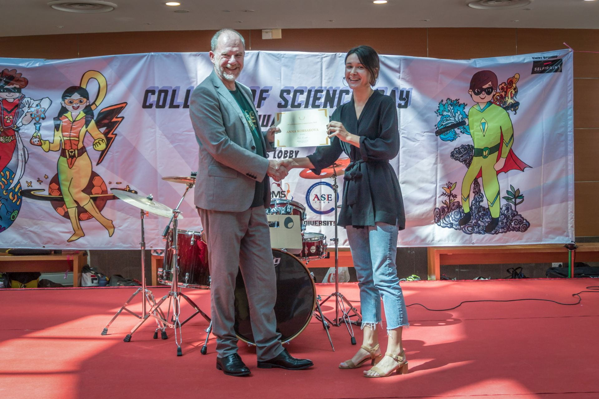
The winners were presented a prize by the Dean of Science, Professor Simon Redfern, at CoS Day 2020.
In First Place, Anna Korsakova from School of Physical & Mathematical Sciences

Chaotic behavior is possible in a variety of complex systems, including neural networks. A feed-forward neural network might exhibit chaotic properties if a feedback loop is added from the output to the input (known as backpropagation method). Behaviors that researchers are usually interested in lay in the output, but if you take a look at the dynamics of the network itself, you’ll sometimes see fascinating many-dimensional chaotic attractors.
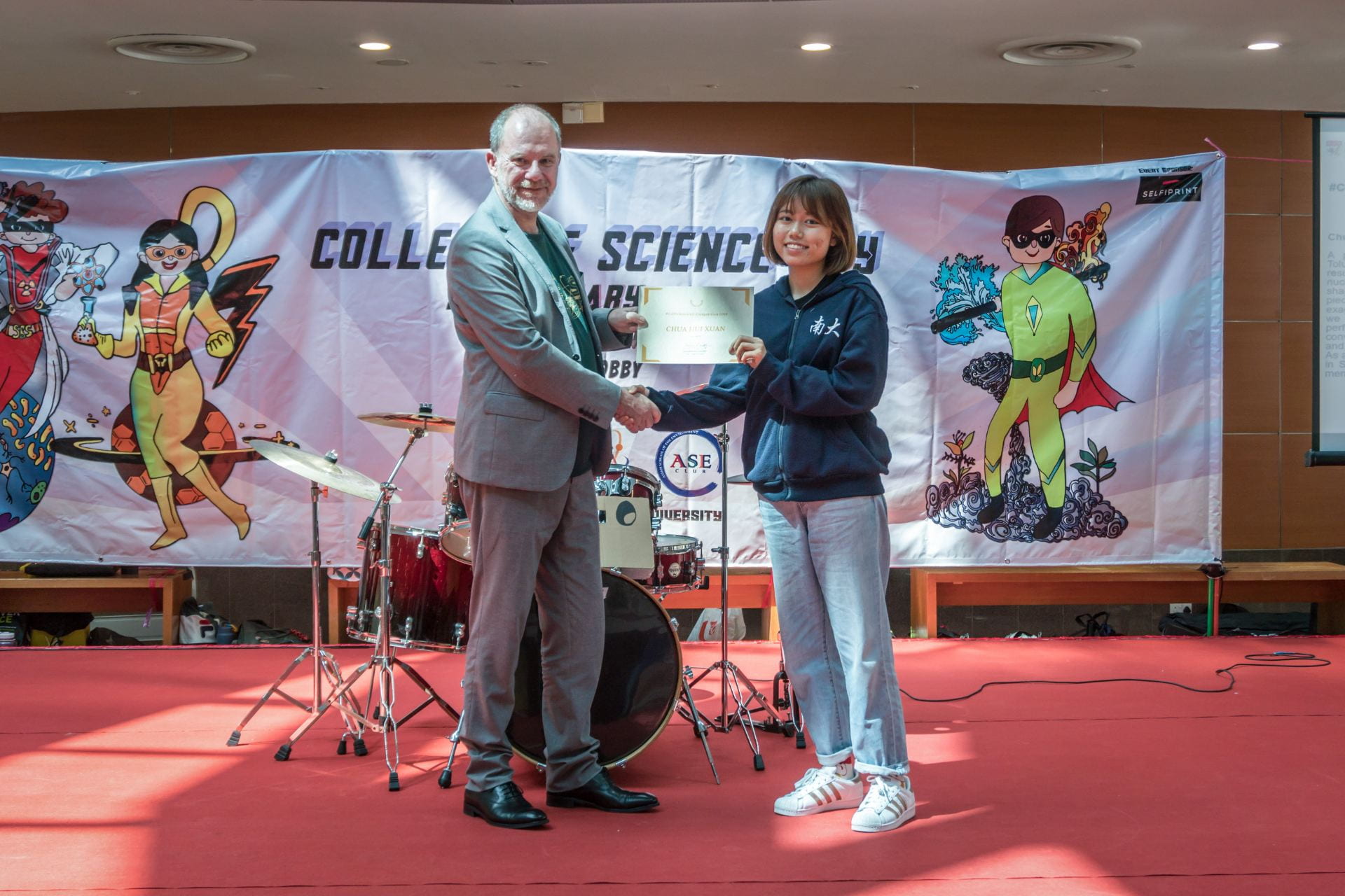
In Second Place, Chua Hui Xuan from School of Biological Sciences

A plant stem sample stained by Toluidine Blue O. A greenish blue result indicates high presence of nucleic acid, while dark blue ovular shapes are likely to be lignin. In this piece, the stained colours have been exaggerated to express the beauty we see and satisfaction felt at a perfect stain. This painting aims to convey the focused mood we enter and the delight we feel at the end. As a reminder of my final lab session in SBS, this was hand-drawn from memory of BS3108’s last lab.
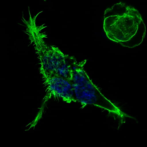
In Third Plaze, Zarina Levitskaya from School of Biological Sciences
Phalloidin is a mushroom toxin used in cell biology for its ability to tightly bind F-actin cytoskeletal protein. Here, tagged to green fluorophore, it allows visualization of a cell under confocal microscope showing how it spreads. Though unable to add much value to my experiment, this image made my day. After looking through numerous cells in a dark room, I saw a bird flying towards sun. What do you see?
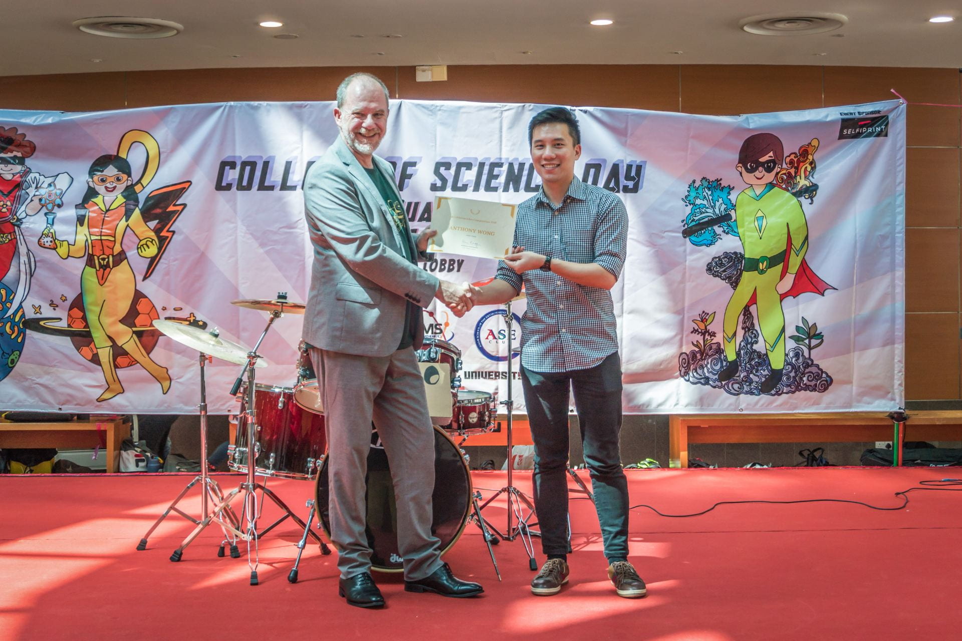
The merit prizes, in no order:
Anthony Wong from Singapore Centre for Environmental Life Sciences Engineering

A microscope image of a fungus called Rhodotorula glutinus. It is a common environmental fungus that can cause opportunistic infections in immunocompromised patients. It has been stained for 18S rRNA (orange) and the nucleus of each cell has been stained with DAPI (blue). This image was taken using a fluorescent microscope.
Liew Woan Feei from School of Biological Sciences

Imagine watching the movie “Joker” the night before, and seeing this in your microscope the next day! This is actually a stained plant cell, assigned to my group as a task to identify the various plant cell structures. With this cell being an unfamiliar one to us, my group tried finding answers on Google. The joker in my group found it under the keywords “scary looking plant cells”…
This plant cell was photographed through a microscope. We first sliced thin layers of the stem of samples provided, then stained them with dyes before viewing them under a light microscope at 40x magnification.

Michael Lodge from School of Physical & Mathematical Sciences

Electrons are quantum mechanical objects characterized by a wavevector k and energy E. Here, electrons moving along the surface of a semimetal encounter an atomic defect that mixes different wavevector states together, elastically scattering the electrons. The result is a standing wave in the electron wavefunction, which is visualized in this scanning tunneling microscopy image. The shape of the standing wave reflects the 4-fold symmetry of this semimetal’s electronic band structure.