Members of the NTU Visual Perception Lab are travelling far and wide to talk about our research.
Arnab presented a poster on “Blink Adaptation for Vergence Eye Movements” at the Vision Sciences Society Annual Meeting in Florida.
Yulia and Maiko are in Rome for the Organization for Human Brain Mapping Meeting, presenting their research on “Perceptual filling-in of the blind spot is not based on activation of monocular region in V1“, and “White Matter Tracts Adjacent to the Cingulate Sulcus Visual Area (CSv) Assessed with Diffusion MRI“, respectively.
Several lab members will be in Osaka at the Asia Pacific Conference on Vision later in July, tp present posters and talks. Egor: “The spatial profile of interocular suppression is modulated by mask speed“. Gerrit: “Motion extrapolation and time compression during eye blinks“.
We hope to meet you, if you’re at any of these meetings!
Hiromasa Takemura from CiNet (Center for Information and Neural Networks) in Osaka will be visiting our lab in February, and give a workshop on Diffusion MRI. Please sign up here!
Date & Time: 12 February 2019 (Tuesday), 2pm – 4pm
Hiromasa Takemura is a tenure-track researcher in the Center for Information and Neural Networks (CiNet), NICT, Japan. Takemura’s research centers on studies of human visual white matter pathways, spanning topics from vision science, comparative neuroanatomy, functional neuroimaging and clinical vision.
Takemura graduated from the University of Tokyo in 2007 with a B.A. in liberal arts. During his Ph.D. at the University of Tokyo, he worked on the psychophysical study of human visual motion perception. From 2012 to 2015, he carried out a series of studies on human visual white matter pathways as a postdoctoral fellow in the Wandell Laboratory at Stanford University. He joined CiNet, NICT in Japan in 2015.
TUTORIAL TALK – 2 pm
White Matter Imaging in Human Neuroscience: Diffusion MRI and Its Applications
In classical studies, neuroanatomists have identified that white matter in the human cerebral cortex is principally composed of bundles of axons (called tracts) and have emphasized the importance of long- range, white matter tracts in the understanding of cognitive functions. Recent advances in diffusion MRI (dMRI) and tractography provides us the opportunity to learn more about white matter tracts in living human brains, and, thus, to better understand how the white matter properties are related to development, disease, learning, and psychological measurements.
In this talk, I will first introduce an overview on the methodologies of acquiring dMRI data. Second, I will discuss several types of methods for modeling dMRI signals in a single voxel from a simpler diffusion tensor model up to complex multi-compartment models. Third, I will introduce advances in modeling white matter tracts (tractography) from deterministic tractography to recent global methods. I will further discuss advantages and limitations of dMRI as a method in systems neuroscience or psychology. The primary target audience is researchers with an interest in using dMRI for human studies. For each topic, background will be provided for audiences without detailed knowledge of specific methods or models. The goal of the tutorial is to provide an overview of what we can and cannot do with dMRI and help audience to consider how it may be useful for their research projects.
SCIENCE TALK – 3 pm
Multi-Dimensional Approaches to Understand the Visual White Matter Pathways
Over the past several decades, vision scientists have made great progress toward the functional organization of the cortical visual system, by identifying numerous brain areas that are specialized into different aspects of visual processing. However, one question remains largely unaddressed: how brain function emerges from either anatomical structures or from anatomical connections across discrete brain areas. Recent progress in diffusion MRI (dMRI) and tractography opens new avenues in understanding the relationship between ana– tomical connections and human visual or cognitive functions. This talk will highlight recent progress in elucidating the relationship between the properties of white matter tracts and visual function by combining dMRI with clinical, behavioral, functional or anatomical measurements.
I will first describe dMRI studies used to identify white matter pathways, such as the vertical occipital fascicles or the stratum proprium of interparietal sulcus, which have been reported in classical anatomical work but have been largely neglected in the literature. I will demonstrate that we can identify these tracts in living humans using dMRI and tractography methods and show that the trajectories of these tracts are consistent with invasive anatomical work performed in non-human primates. Using functional MRI, I will also discuss possible functions of these tracts. I will then describe my recent approaches for interpreting the tissue properties of visual white matter pathways in order to understand retinal disease and human visual performance by comparing dMRI-based measurements with clinical or psychophysical measurements. Finally, I will discuss some controversies concerning visual white matter pathways and how the analysis of ultra-high resolution polarized light imaging (Axer et al., 2011) data will help us to disentangle existing debates and further improve our understanding of visual white matter pathways.
Maiko Uesaki arrived here from Kyoto and will be working on MRI studies of visual areas in the intraparietal sulcus. Egor Ananyev recently finished his PhD at NUS and will be studying interactions between eye blinks and visual attention.
Gerrit answered some questions from the new journal Communications Biology of the Nature family. Read the full interview here: https://doi.org/10.1038/s42003-018-0130-7
Gerrit and Wee Kiat are presenting recent results from our lab at the European Conference on Visual Perception in Trieste, Italy.
Wee Kiat is showing results from our collaboration with the Laboratoire Psychologie de la Perception on Paris on how eye blinks and saccades interact to correct for gaze stability errors:
“Gaze position changes across blinks carry over to saccade landing errors”
Wee Kiat Lau, Thérèse Collins, Gerrit Maus
Gerrit will summarize some of the work going on in our lab during a symposium dedicated to “What are eye blinks good for? Perceptual, oculomotor, and cognitive effects of eye blinks“.
More information can also be found in these two manuscripts that we have just released on PsyArXiv:
The lab, 2018, after managing escape from the “Mausoleum”
After two busy weeks of helping to host and attending several local conferences (PRNI, BrainSTIM, OHBM, NBIA), we took a half day off to attempt an escape from the Mausoleum (we managed… with bare seconds to spare).
Aaron, Wee Kiat, and Yulia staffing the bar at the BrainSTIM reception
Welcome to Arnab Biswas, who joined our lab this week as Lab Manager and Research Assistant. He will be taking care of our equipment and the new lab, and embark on his own research projects soon.
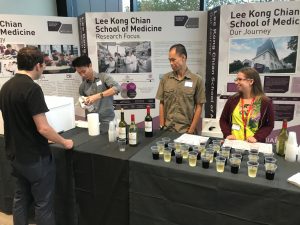
The lab just finished a successful visit to the Vision Science Meeting in Florida. Aaron, Wee Kiat and Yulia all presented their latest research findings as posters, and Christine Veras (now at University of Texas at Dallas) showed her “Silhouette Zoetrope” at the Demo Night.
This week an eminent vision scientist from Germany will visit our lab at NTU. Lothar Spillmann from the University of Freiburg is the discoverer of the watercolour illusion, the co-founder of the European Conference on Visual Perception (ECVP), and has made numerous contributions to the field of Gestalt Psychology, visual perception, and neurophysiology of the visual system.
Lothar will present a CLASS Distinguished Lecture on Tuesday, April 24th at 3 pm in the HSS Conference Room (see details below). All welcome! (Please register here)
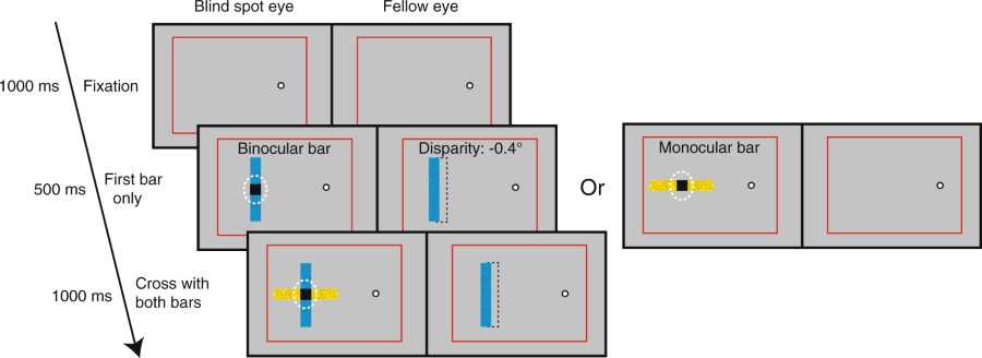
In “Illusory occlusion affects stereoscopic depth perception”, published today in Scientific Reports, we show that filled-in percepts in the blind spot can influence our perception of depth of other objects. When two bars cross in the blind spot–one that is filled in and one that is visible to both eyes with an actual disparity signal that should determine its perceived depth–the filled in bar can nevertheless perceived to be in front and also “push” the other bar to be seen further away. This finding tells us more about filling-in processes occuring in visual cortex and how they can influence other perceptual processes.
Congrats to Mandy Chen from UC Berkeley, who worked on this study together with Rachel Denison (now at NYU), David Whitney, and Gerrit Maus (at NTU Singapore).